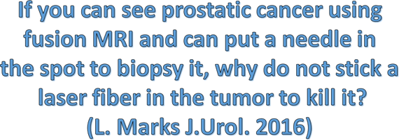
In 2016, visionary American urologist Prof. L. Marks proposed a groundbreaking concept:

What is Echolaser?
Echolaser ablation is a percutaneous, transperineal, ultrasound-guided procedure. Thin optical fibers deliver laser energy for a few minutes, heating the target tissue to induce irreversible necrosis without removing it
Why to use Echolaser?
Advantages over other focal techniques (e.g., HIFU):
- Faster treatment time.
- Lower energy dose.
- Precisely localized effects with clear margins.
- Minimal energy dispersion to surrounding tissues, enabling treatment of critical areas (e.g., near the prostate capsule)
- Preserves erectile function and continence in all cases!
Eligibility for Echolaser Treatment:
- Maximum of two cancerous foci identified via fusion biopsy.
- Gleason score: 6, 7, or 8.
- No capsular infiltration or distant metastases.
Echolaser Treatment
The Echolaser treatment is performed under deep sedation or regional anesthesia. The procedure, which requires no skin incisions, is carried out transperineally and guided by fused Magnetic Resonance Imaging (MRI) and Transrectal Ultrasound (TRUS) images. Laser light is delivered from the source to the targeted tissue through 300-micron optical fibers capable of destroying the tumor.
(From Echolaser in the Treatment of Prostate Cancer – Prof. F. Guercini (Second-Level University Master’s Degree)
The biomedical engineer inputs the tumor data into the system.
The ultrasound probe is inserted, and the laser fiber is activated. Note the grid that serves as a guide during the procedure.
Ongoing treatment. The laser fiber is visible within the tumor (outlined by a green ring). In the upper images, the laser fiber appears white, while in the digital reconstruction of the lower images, the fiber’s trajectory is shown in red. Note that the external grid is reflected on the monitor in the reconstructed view of the prostate and the targeted tissues.
At the end of the treatment, a follow-up MRI confirms that all neoplastic tissue has been ablated.

